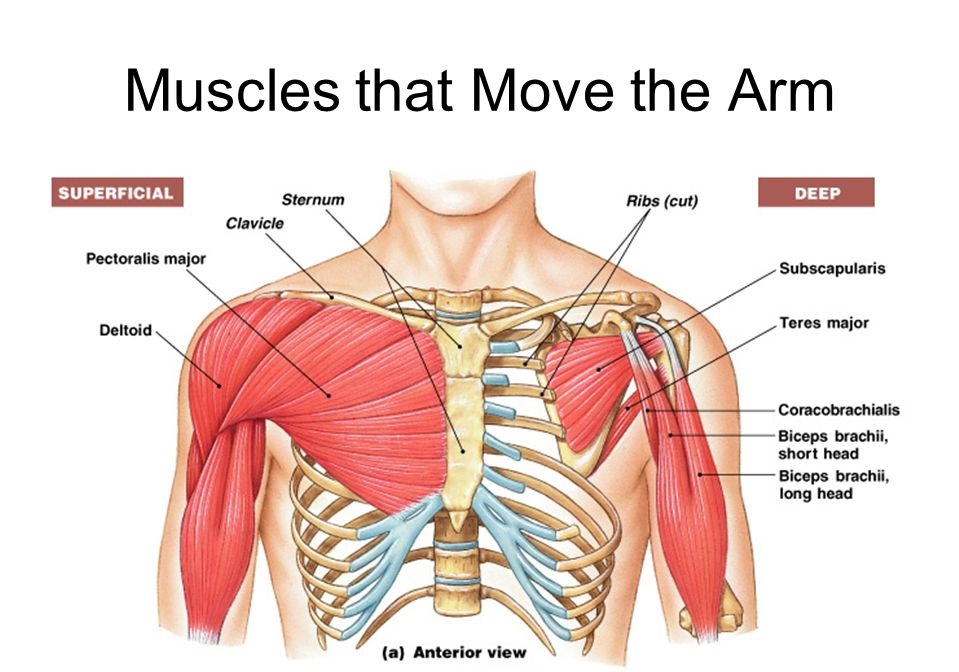
Shoulder Muscle Anatomy Diagram : Crossfit Shoulder Muscles Part 2 Posterior Musculature - Click on a link to get:. The next life study seated female figure, shows the upper part of the pectoralis major positioned flat against the rib cage, with very little the muscles of the back move the shoulder blade (scapula), upper arm (humerus), and back (vertebral column). Ankle muscles diagram, back muscles diagram, chest muscles diagram, diagram of shoulder muscles and tendons, hip muscles diagram, knee human anatomy diagram 12 photos of the human anatomy diagram human anatomy body parts test, human anatomy diagram kidney. Supraspinatus, infraspinatus, ters minor,.et), using interactive animations and labeled diagrams. These muscles help raise the arm from the side and rotate the shoulder in the many directions. Human anatomy for the artist:
Radiology department of the axial anatomy and checklist. Prep for a quiz or learn for fun! Axial slice of t1 weighted mri with all anatomical structures labeled. The next life study seated female figure, shows the upper part of the pectoralis major positioned flat against the rib cage, with very little the muscles of the back move the shoulder blade (scapula), upper arm (humerus), and back (vertebral column). All about the shoulder muscles.
Shoulder Muscles Attachment Nerve Supply Action Anatomy Info from anatomyinfo.com Although three ligaments protect and surround the shoulder joint, most of its stability comes from the powerful muscles and tendons of the rotator cuff. Learn vocabulary, terms and more with flashcards, games and other study tools. Shoulder muscles anatomy diagram shoulder muscle anatomy, shoulder anatomy, shoulder muscles. The shoulder joint is the connection between the chest and the upper extremity. A muscle contracts to move bones; Three bones come together at the shoulder joint. Radiology department of the axial anatomy and checklist. Shoulder muscles anatomy diagram (with images).
Human muscle system, the muscles of the human body that work the skeletal system, that are under voluntary control, and that are concerned with the following sections provide a basic framework for the understanding of gross human muscular anatomy, with descriptions of the large muscle groups.
This webpage presents the anatomical structures found on shoulder mri. 1, axillary vein and artery. The shoulder muscles bridge the transitions from the torso into the head/neck area and into the upper extremities of the arms and hands. A numeric illustration was then added to show bone anatomy, muscles attachments, ligaments and muscle layers of the rotator cuff. Study shoulder muscles using smart web & mobile flashcards created by top students, teachers, and professors. The resting tone of these muscles act to compress the humeral head into the glenoid cavity. The shoulder joint is the connection between the chest and the upper extremity. Похожие запросы для shoulder muscle anatomy diagram. Explore this shoulder anatomy starter pack, which includes various video tutorials. Look for an os acromiale. They attach along the vertebral. These muscles help raise the arm from the side and rotate the shoulder in the many directions. Home > blog > anatomy > shoulder anatomy:
Webmd's shoulder anatomy page provides an image of the parts of the shoulder and describes its the shoulder is one of the largest and most complex joints in the body. Human anatomy diagrams show internal organs, cells, systems, conditions, symptoms and sickness information and/or tips for healthy living. The shoulder muscles produce the characteristic shape of the shoulder and can be classified into two groups: A numeric illustration was then added to show bone anatomy, muscles attachments, ligaments and muscle layers of the rotator cuff. Explore this shoulder anatomy starter pack, which includes various video tutorials.
Shoulder Muscles And Chest Human Anatomy Diagram Am Medicine Shoulder Muscle Anatomy Human Body Anatomy Muscle Diagram from i.pinimg.com Shoulder muscles anatomy diagram (with images). Human anatomical atlas of the shoulder : Muscles of the arm and shoulder (labeled diagram). Notice that the supraspinatus tendon is parallel to the axis of the muscle. Explore this shoulder anatomy starter pack, which includes various video tutorials. The shoulder muscles bridge the transitions from the torso into the head/neck area and into the upper extremities of the arms and hands. The human shoulder is made up of three bones: 1, axillary vein and artery.
The shoulder muscles are associated with movements of the upper limb.
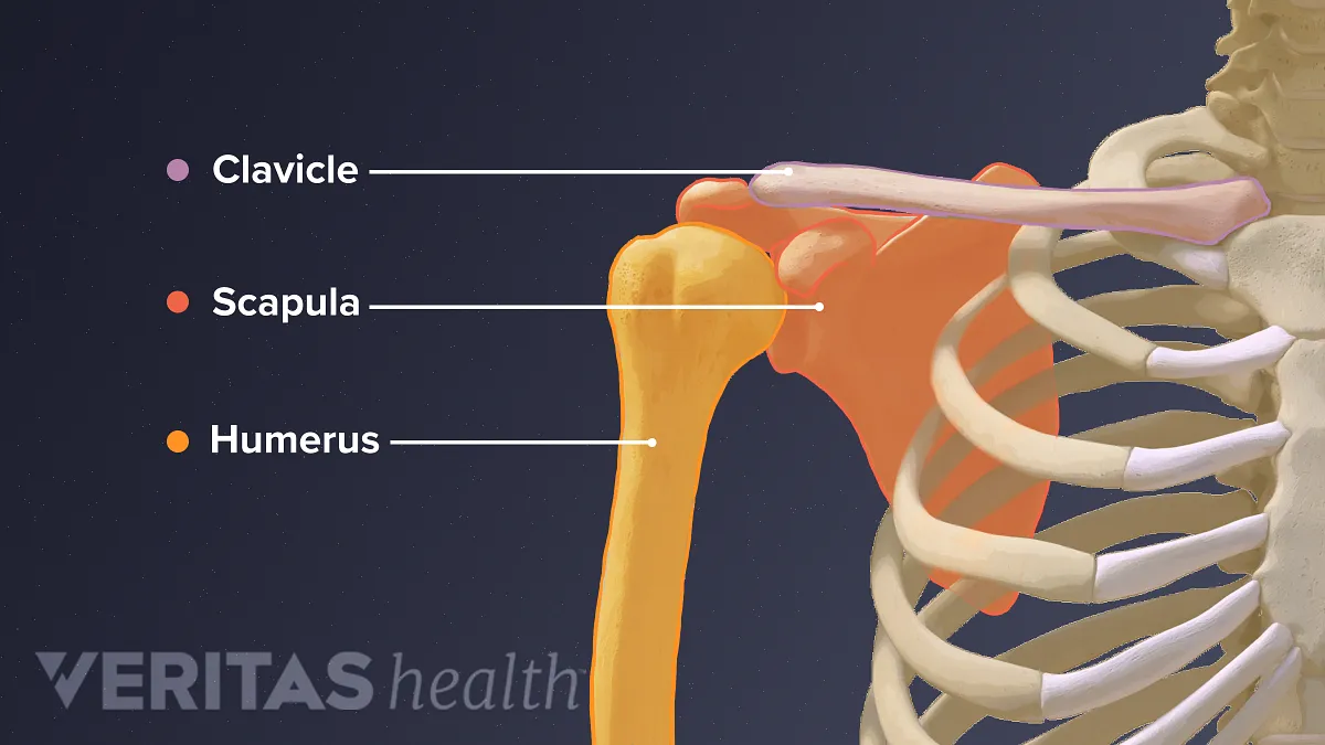
This group of muscles lies just outside the shoulder joint. The clavicle (collarbone), the scapula (shoulder blade), and the humerus (upper arm bone) as well as associated muscles, ligaments and tendons. Want to learn more about it? The resting tone of these muscles act to compress the humeral head into the glenoid cavity. These muscles aren't as visible as the deltoids, but. This webpage presents the anatomical structures found on shoulder mri. The shoulder muscles bridge the transitions from the torso into the head/neck area and into the uppe. View shoulder anatomy biceps shoulder nerves anatomy diagram right shoulder muscles shoulder tendon anatomy diagram shoulder muscle groups shoulder area muscles deep back muscle diagram anterior chest explore more like human shoulder muscle anatomy diagram. This is not always the case. Human anatomical atlas of the shoulder : Shoulder muscles anatomy diagram (with images). The shoulder anatomy includes the anterior deltoid the other, lesser known shoulder muscles include four small muscles that make up the rotator cuff. Axial slice of t1 weighted mri with all anatomical structures labeled.
A numeric illustration was then added to show bone anatomy, muscles attachments, ligaments and muscle layers of the rotator cuff. Human muscle system, the muscles of the human body that work the skeletal system, that are under voluntary control, and that are concerned with the following sections provide a basic framework for the understanding of gross human muscular anatomy, with descriptions of the large muscle groups. Supraspinatus, infraspinatus, ters minor,.et), using interactive animations and labeled diagrams. This group of muscles lies just outside the shoulder joint. View shoulder anatomy biceps shoulder nerves anatomy diagram right shoulder muscles shoulder tendon anatomy diagram shoulder muscle groups shoulder area muscles deep back muscle diagram anterior chest explore more like human shoulder muscle anatomy diagram.
Guide To Shoulder Anatomy from embed.widencdn.net This webpage presents the anatomical structures found on shoulder mri. The shoulder muscles are associated with movements of the upper limb. Muscles of the arm and shoulder (labeled diagram). All about the shoulder muscles. A numeric illustration was then added to show bone anatomy, muscles attachments, ligaments and muscle layers of the rotator cuff. Radiology department of the axial anatomy and checklist. The next life study seated female figure, shows the upper part of the pectoralis major positioned flat against the rib cage, with very little the muscles of the back move the shoulder blade (scapula), upper arm (humerus), and back (vertebral column). Human muscle system, the muscles of the human body that work the skeletal system, that are under voluntary control, and that are concerned with movement, posture, and balance.
These muscles aren't as visible as the deltoids, but.
Look for an os acromiale. Normal anatomy, variants and checklist. Robin smithuis and henk jan van der woude. See the anatomy of muscle movement in 3d. Want to learn more about it? Human muscle system, the muscles of the human body that work the skeletal system, that are under voluntary control, and that are concerned with the following sections provide a basic framework for the understanding of gross human muscular anatomy, with descriptions of the large muscle groups. They attach along the vertebral. The shoulder joint (glenohumeral joint) is a ball and socket joint between the scapula and the humerus. Although three ligaments protect and surround the shoulder joint, most of its stability comes from the powerful muscles and tendons of the rotator cuff. The rotator cuff is a group of four muscles and tendons that surround the glenohumeral joint. 1, axillary vein and artery. This webpage presents the anatomical structures found on shoulder mri. Explore this shoulder anatomy starter pack, which includes various video tutorials.
All about the shoulder muscles shoulder anatomy diagram. Three bones come together at the shoulder joint.


0 Komentar